A field guide
Next stop:
Tanzania
Ewald R. Weibel Auditorium
Which one would you plan for?
Ten million times lighter. And it is not the fly either — the fly is only the delivery system.
The passenger
To the fly, you are a blood meal.
To what the fly is carrying, you are a habitat.
“The fly feeds for thirty seconds and leaves. What it leaves behind stays for years.”
And this is what it is
Trypanosoma brucei
A single-celled parasite, about 25 micrometres long, that lives in your bloodstream in plain sight of your immune system.
“Everything I am going to show you today happens inside this one cell.”
One of everything
genome
Your cells carry hundreds of mitochondria and thousands of copies of the mitochondrial genome. Lose a few and nothing happens. This cell has one of each.
“There is no spare. Every division has to get it right, or the daughter is dead.”
Strip away the biology
and moves while it runs
whatever they do
and must not be dropped
“How do you hold something perfectly still, when the thing holding it never stops moving?”
Three sentences
“Read in order, those three sentences are the model.”
Back where we started
One protein, in one parasite,
holding one genome in place.
The parasite cannot survive without that genome, and it cannot inherit it without this connection. That is why a structure this small is worth four years.
Introduction
It is dangerous
- Sleeping sickness is spread by the tsetse fly
- Affects humans as Human African Trypanosomiasis (HAT)
- Affects live stock as Animal African Trypanosomiasis (AAT)
- Is a Neglected Tropical Disease (NTD) - why?
- WHO: elimination as a public health problem by 2030
Introduction
Milestones in the field of T. brucei
- put the milestonres here as a map
- and the contributions of the Tb field to braoder biology and where to expand the fild in order to better understand the parasite
Introduction
why its important to look at this
- where is the field at this moment and what is the knowledge
A model that allows to define a conceptual problem which is testable
- We have single-copy organelles: flagellum, mitochondrion, kDNA
- Organized subcellular localization of proteins → experiments are reproducible
- Conceptual problem: how to establish a cross-organelle bridge? Flagellum → kDNA
- TAC as a multiprotein complex seems to solve this
- How to test? protein localization allows us to test a function of a protein directly
- Think: possible functions: a physical multiprotein connection connecting organelles can register and possibly integrate information
Overview of the important components
One thing of every(thing)
- One flagellum
- Linked through the Basal Body
- The Tripartite Attachment complex
- One mitochondrion
- Connect to one Kinetoplast DNA (kDNA): the DNA inside the mitochondrion
- Each structure is inherited faithfully without mistakes
- Needs to happen spatially and temporally controlled
Model organism
T. brucei is a model organism
- Subcellular organization is highly ordered and reproducible
- Specifically we have single copies of: the flagellum, the mitochondrion, the kDNA
- Through mapping of each protein we have access to the localization of each protein
- They are easy to culture
- Ideal for conceptual questions on structural biology, specifically in situ
- There are two major difficulties to make the information accessible in situ as well as time resolved
The flagellum
Flagellum, Paraflagellar rod and Flagellar pocket
- Two functions: locomotion and turnover, corkscrew motion
- The paraflagellar rod connects the flagellum to the cell body
- The flagellar pocket is an integration hub to the environment
- Our understanding comes from the field of cell biology and limited structural results
- Knowledge gap: how the flagellar motion is attenuated before it reaches the kDNA
The Tripartite Attachment Complex (TAC)
The Tripartite Attachment Complex (TAC)
- All the components of the TAC are essential - how do we know?
- The TAC consists of 8 proteins, a multiprotein complex
- Assembly in a hierarchical manner (show the hierarchical model here)
- concept: multiple individual structural components build one functional unit - other examples of emergent properties
- Gap: how the individual components appear on a molecular level and how they interact
The mitochondrial genome
The kinetoplast DNA (kDNA)
- Unique to Kinetoplastides: a disk-shaped DNA network located in the mitochondrion
- Minicircles: ~1kb each, encoding guide RNAs
- Maxicircles: ~23 each, encoding rRNAs and respiratory complex subunits
- The kDNA is connected to the basal body via the TAC
- The segregation paradox: six single-copy components: 64 possible segregation outcomes
- Knowledge gap: the forces driving the directional movement of the kDNA
p197
p197: the protein that bridges the gap
- 30-35 repeats, each 175 amino acids, almost identical sequence
- p197 shows a polarity respective of the N- and C-terminal end in relation to the basal body and the outer mitochondrial membrane (show image of N- and C-terminal oriented fluorescence)
- Functionally p197 is a blackbox
- p197 is suggested to have a dual function: ensure the physical connection and structural integrity
- Gap: how the structure or p197 connects to its function as a probably mostly α-helical secondary structure and its intermolecular and potentially intramolecular interaction
Methodology
Bridging scales using Cryo-Electron Tomography and live-cell imaging
- In order to bridge scales we need to increase resolution (make animation, show image or bridging scales of the method and where fl and EM is)
- Fluorescence microscopy allows to look the time domain, but lack molecular resolution
- The introduction of cryoET enables to observe the protein in question in situ
- This means in a near native state which without chemical fixation
- In our case there are alterations: detergent treatment
- Gap: molecular sociology of p197 or any TAC component
Research question
So the question is …
- p197 permanently links the basal body and the kDNA across the cytoplasm and organelles and has never been seen in situ
→ Is p197 a stiff rod or a flexible molecule?
Sample preparation
From the cell to the tomogram
Single particle trials
blabla
Live-cell imaging
Immobilization of cells
Detergent treatment
Making cells transparent to electrons
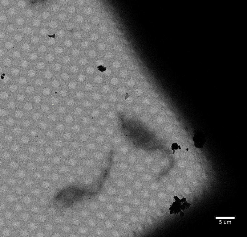
- Mean free path of electrons: inherent limitation to around 500 nm
- This means that untreated T. brucei cells are non-translucent - low-contrast and mostly black
- Goal: alter the sample as little as possible with as much destruction as necessary
- Established protocol for flagellar extraction by Sasse and Gull
- 0.1 % Triton X-100: removes the membranes
- 1 M NaCl + 0.1 % Triton X-100: high salt breaks the ionic contacts between the subpellicular microtubules
Detergent treatment
The possible effect of Triton X-100 and NP-40
- Observation: Triton X-100 renders cells more broken than NP-40
- Both are similar: non-ionic detergents, head group, concentration: 8 mM (35x CMC Triton X100 and 30x CMC NP-40)
- Different: hydrophilic tail → Triton X100: more like a cone
- Possible reason: water-coordination on the hydrophilic tail accounts for the different visual effect and micelle reservoir size does not, but: micelles themselves also act
Triton X-100 n ≅ 9 - 10
NP-40 n ≅ 9
Detergent treatment
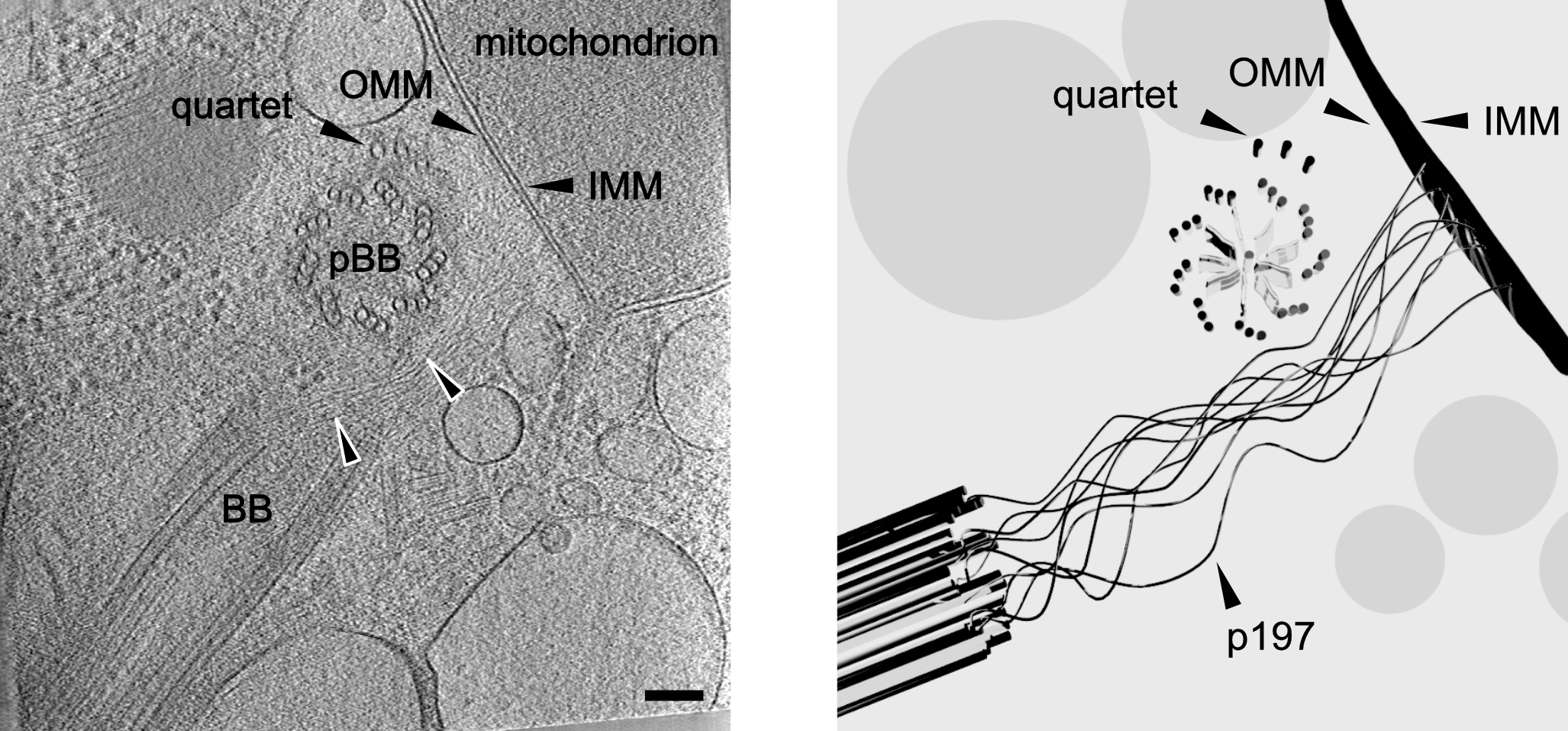
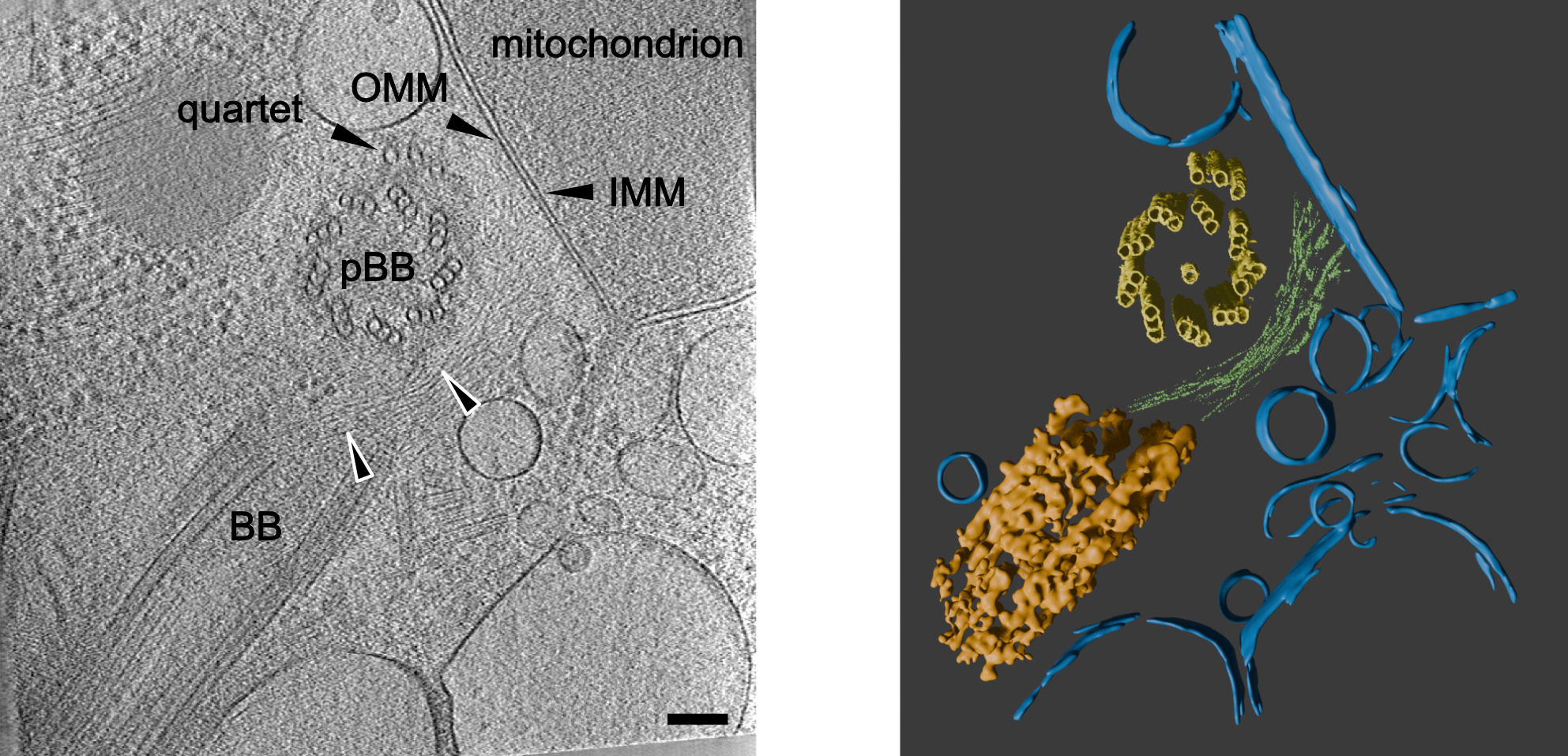
The p197 protein in situ for the first time
Detergent treatment
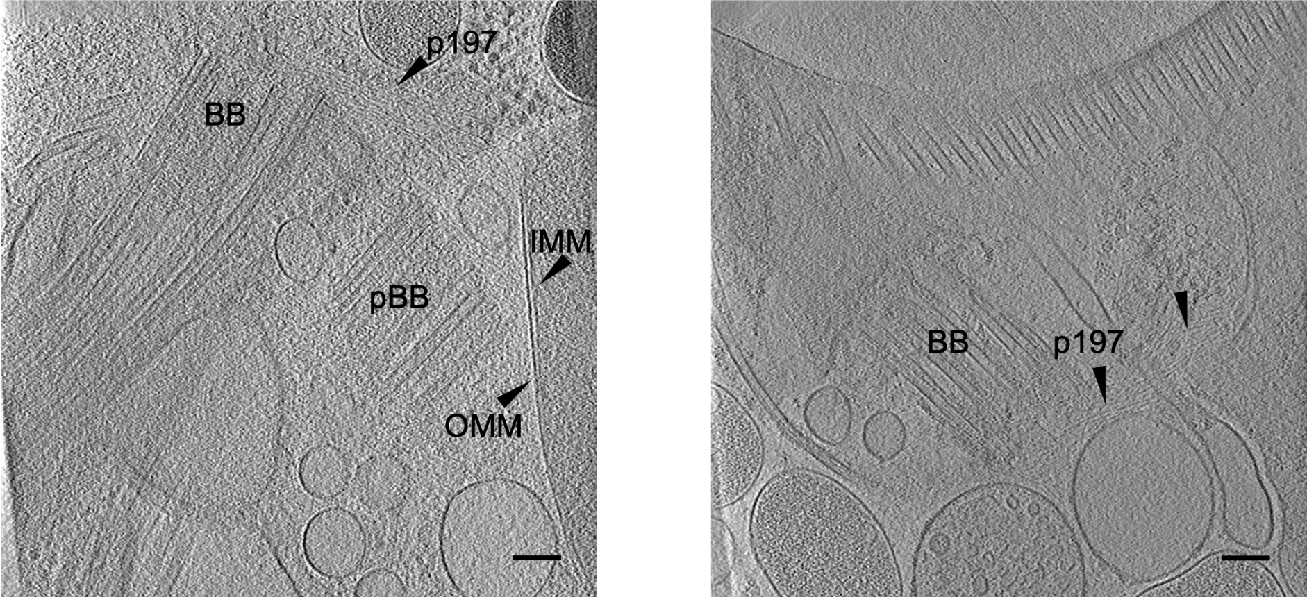
The p197 protein in situ for the first time
Detergent treatment
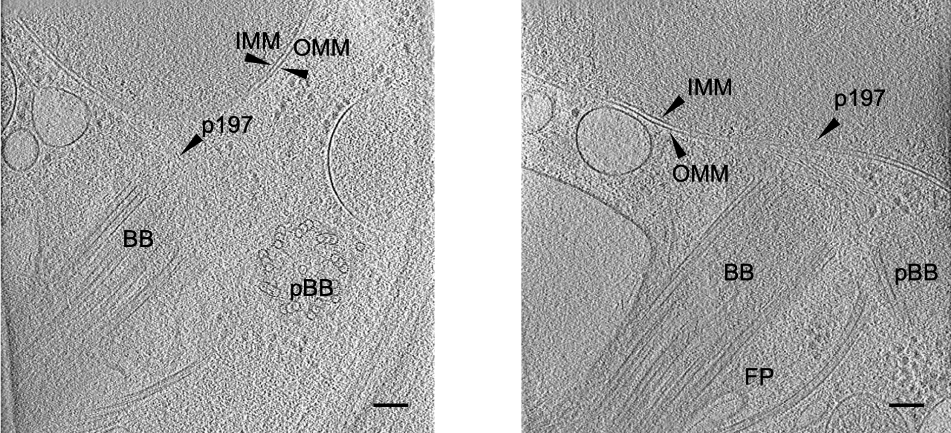
The p197 protein in situ for the first time
Detergent treatment
The p197 protein in situ for the first time
Detergent treatment
The p197 protein in situ for the first time
Detergent treatment
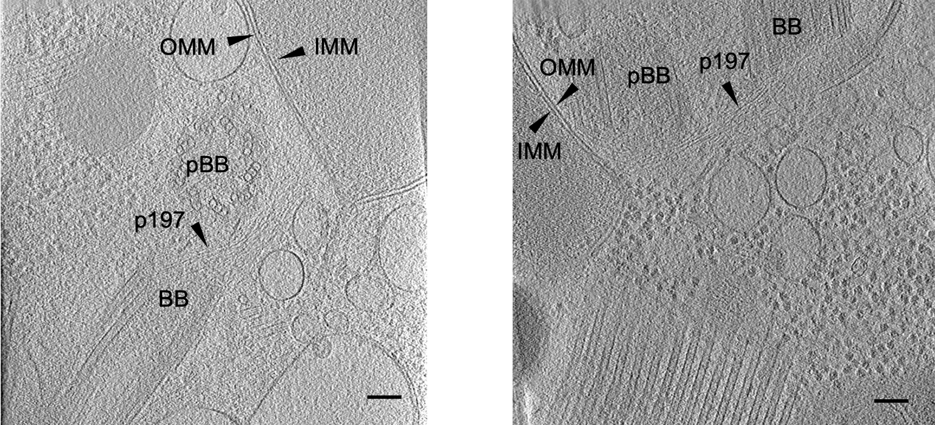
Absence of detergent and flagellar extraction
Detergent treatment
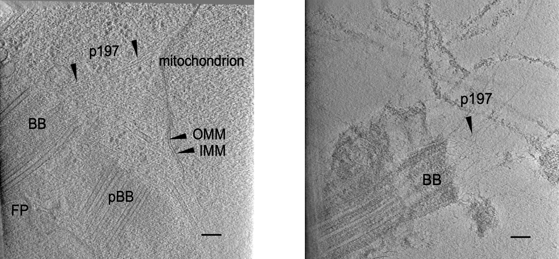
Observations based on differential detergent treatments
- Untreated: mostly opaque, but basal body and pro-basal body are visible
- Triton X-100: cellular content is completely removed, but the C-terminal end of the p197 stays attached to the basal body, however not the N-terminal end
- NP-40: cellular context is undisturbed, C- and N-terminal end are clearly intact and at the expected subcellular location
- Under all three conditions: p197 seems to look the same as a filament that emerges from the basal body
Filament length measurements
Tracing filament in 3D
- Over the time given, ~300 tomograms were recorded, the 12 best looking segmented
- Manual tracing using livewire tool in IMOD
- Every pixel is connected to every neighboring pixel
- Put a low cost if the next pixel is dark, otherwise but a higher cost
- Find the cheapest path between manual annotations
- Observation: visual inspection is necessary and does not work in z-direction but the contour stays connected
Filament length measurements
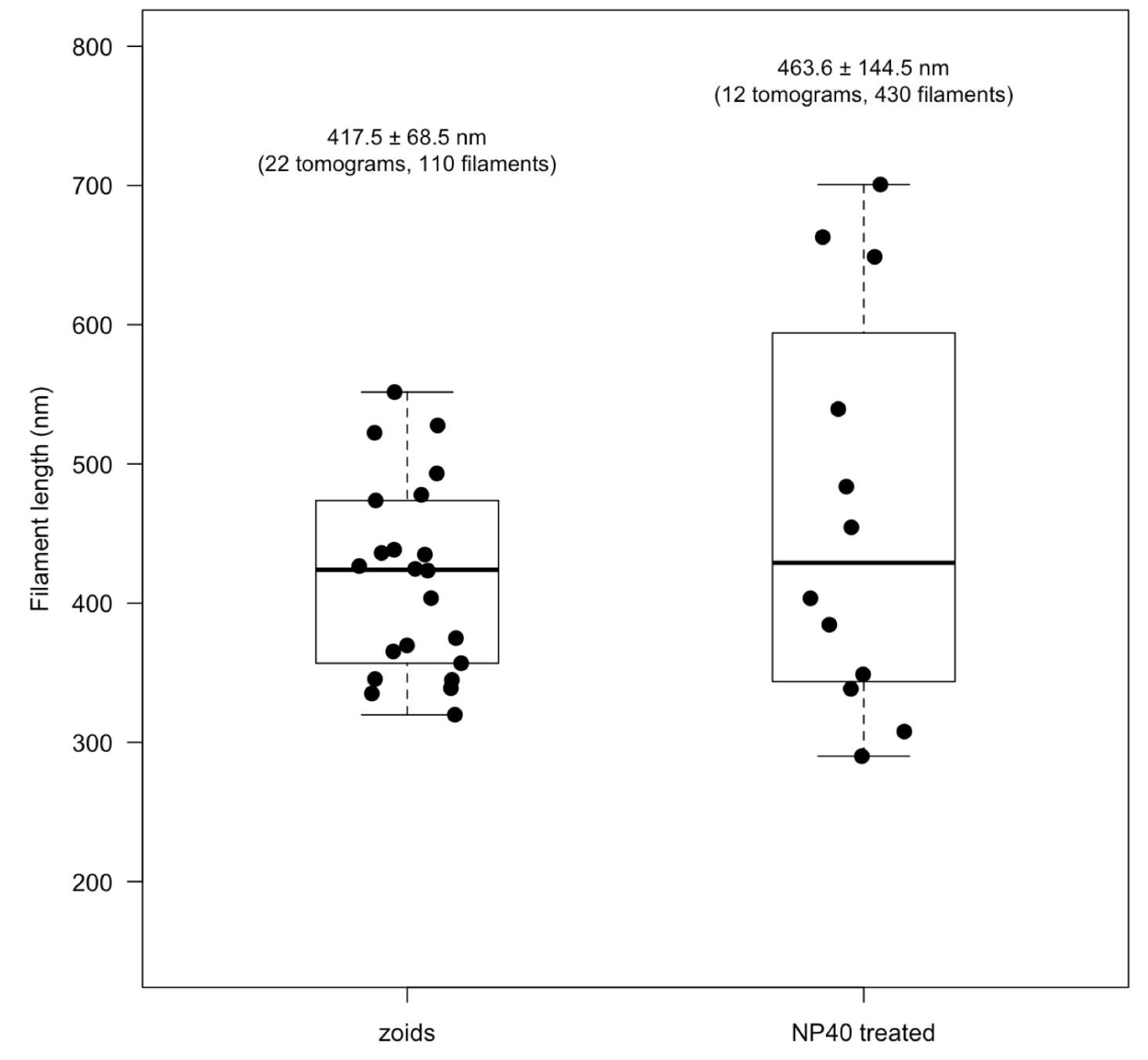
Two independent measurements show similar length
- Left: zoid cells, 417.5 ± 68.5 nm
- Right: wild-type cells, 463.6 ± 144.5 nm
- Observation: wider spread for the NP-40 treated cells: reason: every visibly distinguishable filament was measured whereas on zoids five per tomogram were traced
Filament length measurements
p197 does not have a fixed length
- From Aeschlimann et al.: 2D distance between basal body and outer mitochondrial membrane is 270 nm
- This is based on expansion microscopy where the cells are chemically fixed
- The measurements comes from a point-to-point path
- The gap between the basal body in cryoET is even shorter: ~145 nm
- Tracing in 3D: 417 nm (zoids) and 464 nm (wild-type)
- Apparent contradiction in length, however different measurements: point-to-point vs. filament path
- Apparent contradiction and no falsification of the hypothesis
Live-cell imaging
Immobilized cells are still swimming (normally)
- Fluorescent proxy-measurement for movement across the TAC: basal body and kDNA labelled
- ~100 movies were recorded, nine cells were tracked over 800 frames each
- Using TrackMate the flagellar beat frequency was determined: 8.43 ± 0.48 Hz
- In agreement with 3D tracking of swimming cells: 6.9 ± 2.2 Hz
- Comparison is valid because a reduction in dimensionality does not affect the temporal frequency
Live-cell imaging
Tracking the two anchor points of p197
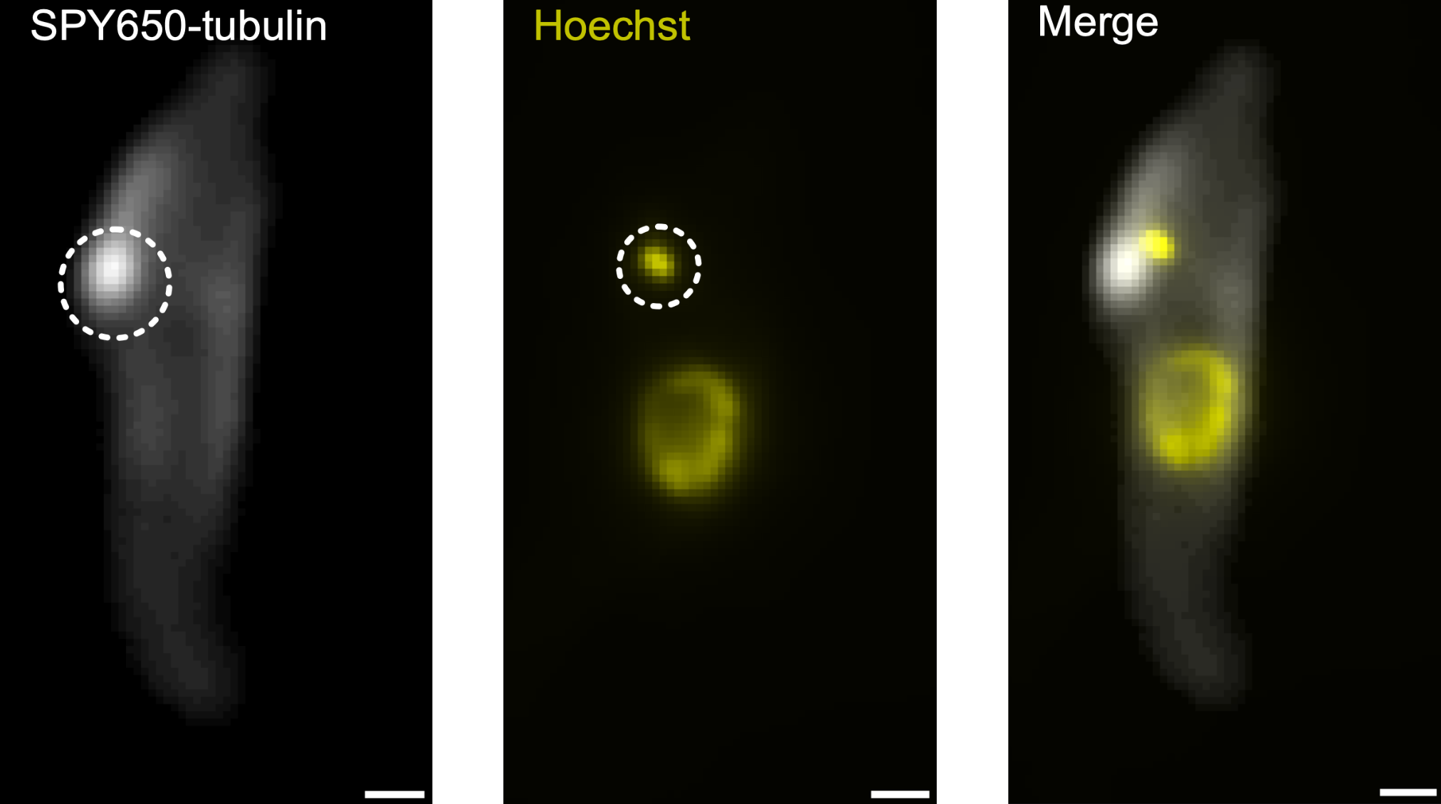
- SPY650-tubulin: stains microtubules and the basal body
- Hoechst: stains the DNA
- Both channels are recorded per cell
- Frequency was measured by centroid tracking using TrackMate
- Right: circles show the tracked regions
- Observation: the new immobilization method works - but can we quantify this movement?
Live-cell imaging
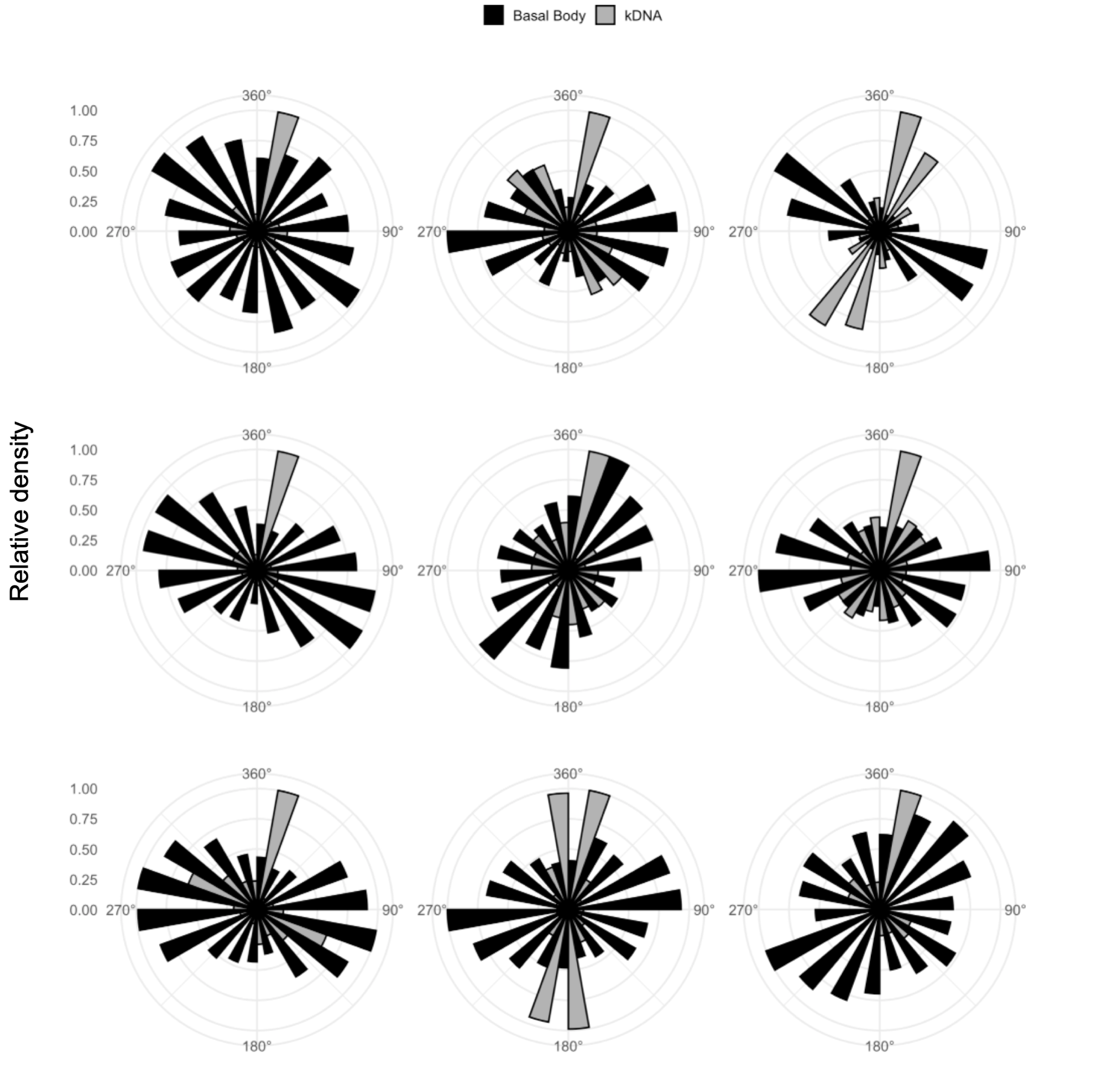
The basal body moves more than the kDNA
- Rose plots: relative density as a function of displacement angle
- Basal body displacement is in all directions while the kDNA has narrow directional movement
- Observation: the two anchor points of the p197 move in different directions
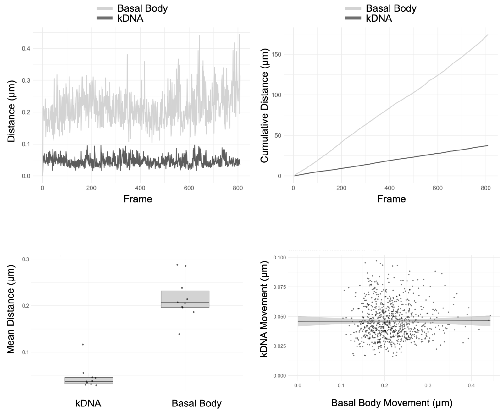
Live-cell imaging
The basal body and the kDNA move independently
- Top left: the basal body moves more than the kDNA
- Top right: the sum over the movie, ~160 μm for the basal body and ~35 μm for the kDNA
- Bottom left: the values per cell and movie: basal body 0.216 ± 0.052 µm and kDNA 0.046 ± 0.015 µm
- Bottom right: can the movement of the basal body predict the movement of the kDNA
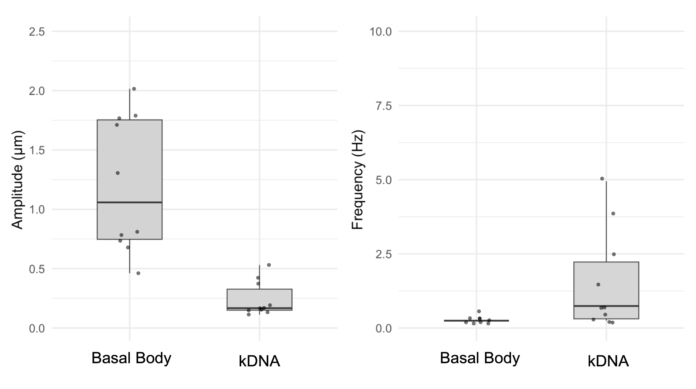
Live-cell imaging
The basal body movement is absorbed by the TAC
- FFT analysis of both tracked signals
- Amplitude basal body: 1.44 ± 0.64 μm
- Amplitude kDNA: 0.33 ± 0.25 μm
- Absorbed difference: 1.11 μm (p = 0.00247)
- Frequency: difference is not significant (p = 0.0629)
- Observation: the movement is reduced at the kDNA
Live-cell imaging
Conclusion from the live-cell experiments
- The basal body moves 4-5 times more than the kDNA
- We cannot predict the movement of the kDNA based on he movement of the basal body
- This means: the basal body and the kDNA move independently
- → the TAC somehow mitigates the flagellar movement
- Possible culprit: p197
Experimental observation summary
What the cryoET and the live-cell imaging tells us
Cryo-Electron Tomography
- We successfully visualized p197 for the first time in situ
- The anchorage at the N- and C-terminal seems to be different in stability
- The lengths of the filaments measure are about three times the gap between the basal body and the outer mitochondrial membrane
- Filament length varies, even at one basal body
Live-cell imaging
- Immobilized cells beat at a frequency comparable to reported values
- The basal body moves four times more than the kDNA
- The two move independently
→ p197 is a slack rope and the flagellar movement is attenuated between the basal body and the kDNA
Integration of experimental observation
The buffer model
- The basal body moves 4–5× more than the kDNA
- Basal body and kDNA move independently
- p197 is stable upon detergent treatment
- p197 filament length is heterogeneous
Limitation of the buffer model
Limitations and ideas to put new boundaries
Limitations
- Presence of detergent might alter the TAC structure in situ
- The live-cell 2D tracking is a reduction in dimensionality
- Tracking is limited to the anchor points, i.e. the N- and the C-terminal end of p197 and not p197 itself
- The p197 as the main culprit of attenuation cannot be isolated
- The noise level of a non-moving object needs to be assessed properly
Further experiments
- Visualization of the TAC components and compare to our expectation
- Subtomogram averaging of p197 as well as all the TAC components
- Live-cell imaging and tracking in 3D, this is partially achieved using CyGEL
- in CyGEL the cells stay alive for more than 24 h and can be recovered and re-cultured
Take-home message
Is p197 stiff or flexible?
- p197 is long, stable (at least to some degree)
- p197 has not one fixed length and bridges a bigger than necessary gap, the sequencing data is misleading
- The flagellar beating is absorbed between the basal body and the kDNA
- TAC is not only a physical bridge between two organelles but seems to have additional function in attenuating the flagellar beating
Acknowledgements
- Ioan Iacovache
- David Kalbermatter and Marek Kaminek
- Prof. Benoît Zuber
- Prof. Torsten Ochsenreiter
- Prof. Thomas Lemmin, Prof. Sebastian Leidel and Prof. Paul Guichard
- Prof. Simone Mattei and Georg Wolff @ EMBL
- Reto Caldelari and Yury Belyaev @ MIC, Kodzo Atchou
- CEM curriculum
- Prof. Ernst Hunziker
- Adolfo Odriozola and Jana Leuenberger